
How 3D Imaging Makes Dental Implant Surgery Safer and More Accurate
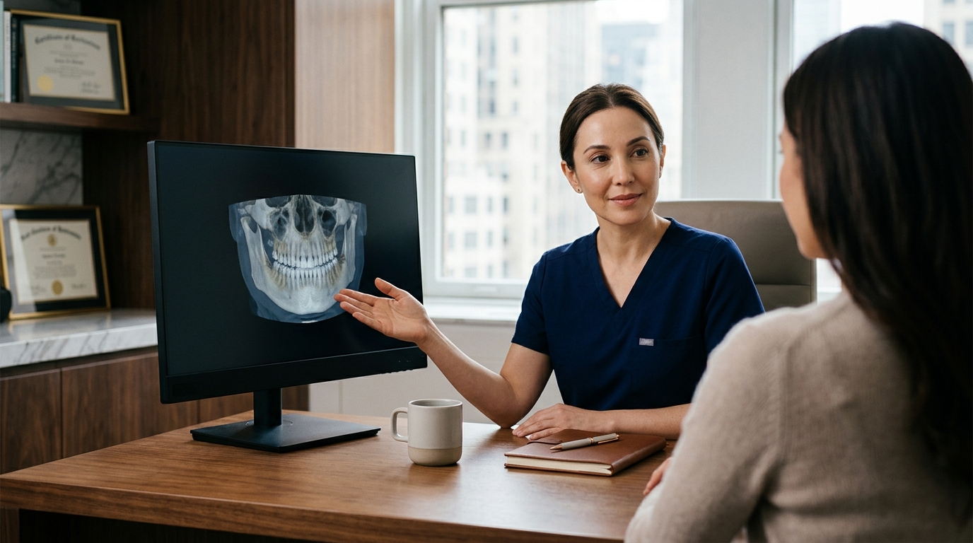
3D imaging technology makes dental implant surgery safer and more accurate by providing a detailed, three-dimensional view of the jawbone, nerves, and sinuses before any incision is made. At Park Avenue Oral, Maxillofacial & Implant Surgery, we use Cone Beam Computed Tomography (CBCT) scans to map your unique anatomy. This digital blueprint allows our surgeons to plan the exact position, angle, and depth of each implant, significantly reducing the risk of complications and ensuring a stable, long-lasting result.
The Limitations of 2D X-Rays in Implant Planning
Traditional dental X-rays provide a two-dimensional view of the mouth, which can be misleading when planning complex procedures. A 2D image flattens three-dimensional structures, making it difficult to accurately measure bone height or determine the precise location of vital structures like the inferior alveolar nerve. This lack of depth perception can lead to underestimating the amount of bone available for an implant. according to Magnetic resonance imaging
When a surgeon relies solely on 2D imaging, there is a higher risk of placing an implant too close to a nerve or sinus cavity. This can result in post-operative pain, numbness, or the need for additional corrective surgeries. By recognizing these limitations, we prioritize 3D imaging to eliminate the guesswork associated with traditional radiographs.
Why Depth Matters
Depth perception is critical for assessing bone volume. Without it, a surgeon cannot accurately determine if a sinus lift or bone graft is necessary before the procedure begins. This lack of clarity often leads to unexpected changes in the treatment plan during surgery, which can increase procedure time and patient discomfort.
How CBCT Scans Work
Cone Beam Computed Tomography (CBCT) is a specialized form of 3D imaging that captures detailed cross-sectional images of the head and neck. Unlike medical CT scans, CBCT uses a cone-shaped beam of X-rays to create a high-resolution 3D model of the jaw with lower radiation exposure. This technology allows us to rotate and zoom in on specific areas of the jaw to evaluate bone density and quality.
At Park Avenue Oral, Maxillofacial & Implant Surgery, the CBCT scan is the foundation of our digital workflow. The scan provides a comprehensive view of the alveolar ridge, allowing us to measure the exact width and height of the bone. This data is essential for determining if the bone is sufficient to support an implant or if augmentation is required. The precision of CBCT ensures that our surgical plans are based on accurate anatomical data rather than estimates. according to 3D Guided Dental
Low Radiation, High Detail
CBCT technology is designed to minimize radiation exposure while maximizing image clarity. The scan is quick and non-invasive, typically taking only a few seconds to complete. This makes it a safe and efficient tool for pre-surgical planning, providing the necessary detail without subjecting the patient to excessive radiation.
Virtual Planning and Surgical Guides
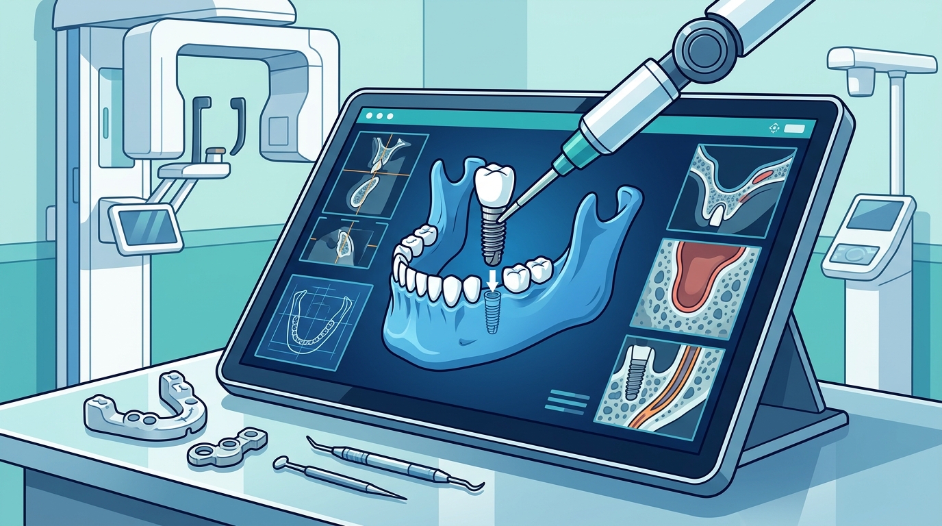
Once the CBCT data is acquired, we use specialized software to create a virtual model of your jaw. This digital environment allows the surgeon to simulate the implant placement before entering the operating room. We can adjust the position, angle, and depth of the implant virtually to ensure it fits perfectly within the available bone. This process, known as Virtual Surgical Planning (VSP), is a critical step in achieving predictable outcomes.
From this virtual plan, we can fabricate a 3D-printed surgical guide. This physical template is placed in the mouth during surgery to direct the drill to the exact location planned on the computer. The guide ensures that the implant is placed with millimeter-level accuracy, reducing the risk of damaging surrounding structures. This level of precision is particularly important for complex cases involving multiple implants or full-arch restorations.
The Role of 3D Printing
3D printing technology allows us to create custom surgical guides and splints that fit the patient's anatomy perfectly. These guides are designed to be comfortable and easy to use, ensuring that the surgical team can follow the plan with confidence. The integration of 3D printing into our workflow enhances the consistency and quality of our surgical outcomes.
Reducing Complications and Improving Outcomes
The use of 3D imaging and guided surgery has been shown to reduce the incidence of complications such as nerve injury and sinus perforation. By visualizing the exact location of these structures, the surgeon can avoid them entirely. This proactive approach to safety minimizes the risk of post-operative issues and promotes faster healing. Patients benefit from a more predictable recovery process with less pain and swelling.
Furthermore, 3D imaging allows for the early detection of anatomical variations that might not be apparent on 2D X-rays. This includes identifying narrow bone ridges or unusual nerve pathways. By addressing these factors during the planning phase, we can design a treatment plan that maximizes the long-term success of the implant. The goal is to provide a safe, efficient, and effective solution for replacing missing teeth.
Long-Term Stability
Accurate implant placement is crucial for the long-term stability of the restoration. When an implant is positioned optimally, it distributes chewing forces evenly, reducing the risk of mechanical failure. 3D imaging ensures that the implant is placed in the strongest part of the bone, providing a solid foundation for the crown or bridge. This attention to detail contributes to the durability and function of the final restoration.
The Park Avenue Approach to Precision
At Park Avenue Oral, Maxillofacial & Implant Surgery, we combine advanced technology with surgical expertise to deliver the highest standard of care. Our team uses CBCT imaging and Virtual Surgical Planning to ensure that every implant placement is precise and safe. We are committed to using the latest tools and techniques to minimize invasiveness and maximize patient comfort. This approach reflects our dedication to providing excellent care in a friendly and caring environment.
Our surgeons are board-certified and fellowship-trained, bringing a wealth of experience to the use of 3D imaging. We understand that technology is only as good as the hands that use it. By integrating digital planning into our clinical practice, we ensure that the technology serves the patient's best interests. Our goal is to provide a seamless and confident experience from consultation to final restoration.
Comprehensive Care
We offer a range of implant solutions, from conventional implants to complex full-arch restorations. Each case is evaluated using 3D imaging to determine the best approach. Whether you need a single tooth replacement or a full smile makeover, our digital workflow ensures that the plan is tailored to your unique anatomy. This personalized approach is a hallmark of our practice.
Key Takeaways
- CBCT scans provide a 3D view of the jaw, offering superior depth perception compared to 2D X-rays.
- Virtual Surgical Planning allows surgeons to simulate implant placement before the procedure begins.
- 3D-printed surgical guides ensure that implants are placed with millimeter-level accuracy.
- 3D imaging helps avoid vital structures like nerves and sinuses, reducing the risk of complications.
- Accurate placement contributes to the long-term stability and success of dental implants.
- At Park Avenue Oral, Maxillofacial & Implant Surgery, we use advanced technology to enhance safety and predictability.
Frequently Asked Questions
Is a CBCT scan painful?
No, a CBCT scan is completely painless and non-invasive. It is similar to a standard dental X-ray but provides a 3D image. The scan takes only a few seconds to complete.
How much radiation does a CBCT scan involve?
CBCT scans use a lower dose of radiation compared to traditional medical CT scans. The radiation exposure is minimal and considered safe for pre-surgical planning.
Do I always need a 3D scan for an implant?
While not every case requires a 3D scan, it is highly recommended for most implant procedures. It provides the detailed information needed for accurate planning and safe placement.
Can 3D imaging detect bone loss?
Yes, 3D imaging can accurately measure bone volume and density. This helps determine if a bone graft is needed to support the implant.
How does 3D printing help in implant surgery?
3D printing is used to create custom surgical guides that fit the patient's mouth. These guides direct the drill to the exact location planned in the virtual model, ensuring precision.
What are the benefits of guided implant surgery?
Guided surgery reduces procedure time, minimizes invasiveness, and lowers the risk of complications. It leads to more predictable outcomes and faster recovery.
Conclusion
3D imaging technology has transformed dental implant surgery by enhancing safety and accuracy. By using CBCT scans and Virtual Surgical Planning, we can map your anatomy precisely and plan your treatment with confidence. At Park Avenue Oral, Maxillofacial & Implant Surgery, we are dedicated to leveraging these advanced tools to provide you with the best possible care. If you are considering dental implants, schedule a consultation with our team to learn more about how 3D imaging can benefit your treatment.